
Stress distribution on the L1/L2 endplates under multiaxial loads: A finite element study
Article Sidebar
Main Article Content
Abstract
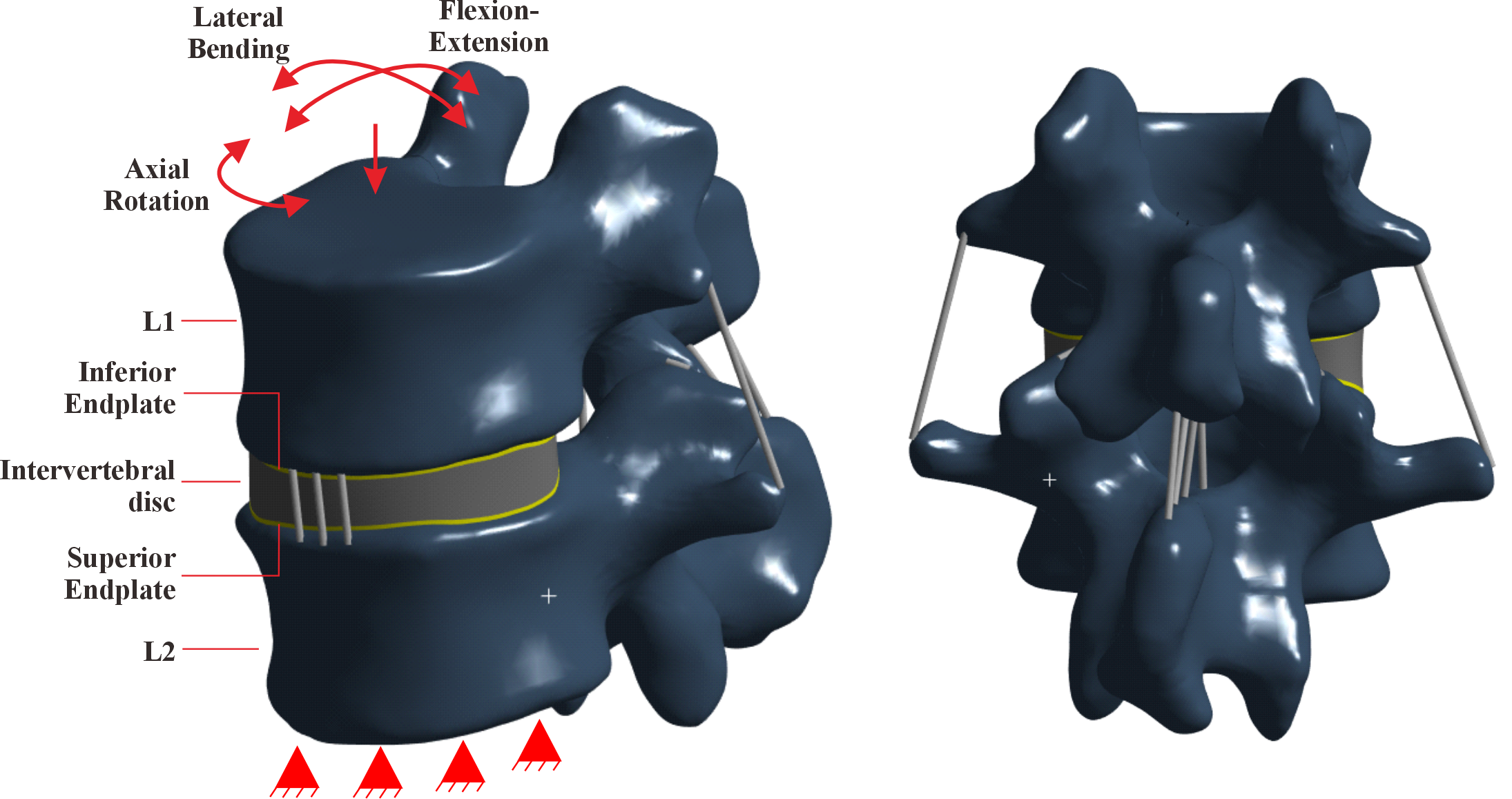
Understanding stress distribution on lumbar vertebral endplates is essential for predicting mechanical failure and guiding clinical interventions. Therefore, this study aims to investigate the von Mises stress patterns on the L1/L2 endplates under multiaxial loading using a 3-dimensional finite element (FE) model derived from CT imaging of a healthy 55-year-old male. Anatomical structures were reconstructed in Mimics 21.0, and simulations were conducted in ANSYS Workbench 2023 R2. Material properties for cortical bone, cancellous bone, and intervertebral disc were assigned based on validated biomechanical data. A compressive load of 500 N and multiaxial moments ranging from 2.5 to 10 N•m were applied to simulate physiological movements, while the inferior surface of L2 was fully constrained to reflect realistic boundary conditions. The results showed that the superior endplate experienced the highest von Mises stress, particularly during flexion and lateral bending, indicating increased vulnerability to mechanical overload. Extension loading significantly reduced stress on both endplates, with a 60.54% decrease on the superior endplate and 69.17% on the inferior endplate. Stress distribution was asymmetrical and was influenced by anatomical features, such as cortical thickness and trabecular alignment. These results show the superior endplate as a biomechanically critical region prone to degeneration, emphasizing its importance in implant design, preventive strategies, and risk assessment for microfracture in high-risk populations.
Downloads
Article Details

This work is licensed under a Creative Commons Attribution-NonCommercial 4.0 International License.
References
[2] M. I. Ammarullah et al., “A review of enhanced total hip prosthesis design and material bearing combination to accommodate Muslim prayer (Salat) movements: Biomechanical, biotribological, and biological perspectives,” Tribology International, vol. 205, no. January, p. 110518, 2025, doi: 10.1016/j.triboint.2025.110518.
[3] W. M. Park, K. Kim, and Y. H. Kim, “Effects of degenerated intervertebral discs on intersegmental rotations, intradiscal pressures, and facet joint forces of the whole lumbar spine,” Computers in Biology and Medicine, vol. 43, no. 9, pp. 1234–1240, 2013, doi: 10.1016/j.compbiomed.2013.06.011.
[4] F. Galbusera and T. Bassani, “Lumbar spine biomechanics: A review of in vitro and computational studies,” Journal of Biomechanics, vol. 94, pp. 73–83, 2019, doi: 10.1016/j.jbiomech.2019.07.004.
[5] Y. X. J. Wáng and others, “Osteoporotic vertebral endplate and cortex fractures: A pictorial review,” Journal of Orthopaedic Translation, pp. 35–49, 2018, doi: 10.1016/j.jot.2018.08.004.
[6] R. Tsujimoto, H. Yamada, T. Nakazawa, T. Sugiura, and H. Ito, “Degenerative disc disease and osteophyte formation in lumbar spondylosis: A radiological and pathological correlation,” Spine Journal, vol. 16, no. 5, pp. 679–687, 2016, doi: 10.1016/j.spinee.2016.01.012.
[7] I. Yamamoto, M. M. Panjabi, T. Crisco, and T. Oxland, “Three-dimensional movements of the whole lumbar spine and lumbosacral joint.,” Spine, vol. 14, no. 11, pp. 1256–1260, Nov. 1989, doi: 10.1097/00007632-198911000-00020.
[8] R. Ismail et al., “Design, fabrication, and performance testing of an energy storage and return (ESAR) foot prosthesis made of prepreg carbon composite,” Mechanical Engineering for Society and Industry, vol. 5, no. 1, pp. 20–32, Jan. 2025, doi: 10.31603/mesi.12652.
[9] H. Li and Z. Wang, “Intervertebral disc biomechanical analysis using the finite element modeling based on medical images,” Computerized medical imaging and graphics, vol. 30, no. 6–7, pp. 363–370, 2006.
[10] A. Zulkifli, A. K. Ariffin, and M. M. Rahman, “Probabilistic finite element analysis of vertebrae of the lumbar spine under hyperextension loading,” International Journal of Automotive and Mechanical Engineering, vol. 3, no. 1, pp. 256–264, 2011, doi: 10.15282/ijame.3.2011.3.0022.
[11] J. M. Daniels, C. Arguelles, C. Gleason, and W. H. Dixon, “Back Injuries,” Primary Care - Clinics in Office Practice, vol. 47, no. 1, pp. 147–164, 2020, doi: 10.1016/j.pop.2019.10.008.
[12] S. Kang et al., “Analysis of the physiological load on lumbar vertebrae in patients with osteoporosis: a finite-element study,” Scientific Reports, vol. 12, no. 1, pp. 1–14, 2022, doi: 10.1038/s41598-022-15241-3.
[13] Z. Shi, J. Liu, X. Yu, L. Jiang, H. Wu, and Q. Pang, “The biomechanical effects of graded upper articular process arthroplasty on lumbar spine: A finite element study,” Journal of Orthopaedic Science, vol. 25, no. 5, pp. 793–799, 2020, doi: 10.1016/j.jos.2019.10.012.
[14] M. Xu, J. Yang, I. H. Lieberman, and R. Haddas, “Lumbar spine finite element model for healthy subjects: development and validation,” Computer Methods in Biomechanics and Biomedical Engineering, vol. 20, no. 1, pp. 1–15, 2017, doi: 10.1080/10255842.2016.1193596.
[15] K. B. Crump et al., “Cartilaginous endplates: A comprehensive review on a neglected structure in intervertebral disc research,” JOR Spine, vol. 6, no. 4, pp. 1–22, 2023, doi: 10.1002/jsp2.1294.
[16] M. Stańczak, M. Biały, and M. Hagner-Derengowska, “Ligament Cell Biology: Effect of Mechanical Loading,” Cellular physiology and biochemistry : international journal of experimental cellular physiology, biochemistry, and pharmacology, vol. 59, no. 2, pp. 252–295, 2025, doi: 10.33594/000000773.
[17] Y. O. Özkılıç, “The capacities of thin plated stiffened T-stubs,” Journal of Constructional Steel Research, vol. 186, 2021, doi: 10.1016/j.jcsr.2021.106912.
[18] J.-P. Roux et al., “Contribution of trabecular and cortical components to biomechanical behavior of human vertebrae: An ex vivo study,” Journal of Bone and Mineral Research, vol. 25, no. 2, pp. 356–361, 2010, doi: 10.1359/jbmr.090803.
[19] U. K. Ezemagu, F. Akpuaka, E. Iyidobi, and C. Anibeze, “Considering the surface area and sagittal angle in a pair of lumbosacral facets: Determining the structural relevance of asymmetric facets at the lumbosacral junction,” Journal of the Anatomical Society of India, vol. 69, no. 4, pp. 237–242, 2020, doi: 10.4103/JASI.JASI_53_19.
[20] Y. Wang, H. Wang, F. Lv, X. Ma, X. Xia, and J. Jiang, “Asymmetry between the superior and inferior endplates is a risk factor for lumbar disc degeneration,” Journal of Orthopaedic Research, vol. 36, no. 9, pp. 2469–2475, 2018, doi: 10.1002/jor.23906.
[21] V. Boddapati, F. Yuk, and S. Virk, “A Cadaveric Comparison of Discectomy Performance During Transforaminal Lumbar Interbody Fusion Approach Using an Endoscopic Technique Versus a Minimally Invasive Tubular Approach,” Spine, vol. 50, no. 10, pp. 713–719, 2025, doi: 10.1097/BRS.0000000000005122.
[22] L. Zhang, H.-M. Li, R. Zhang, H. Zhang, and C.-L. Shen, “Biomechanical Changes of Adjacent and Fixed Segments Through Cortical Bone Trajectory Screw Fixation versus Traditional Trajectory Screw Fixation in the Lumbar Spine: A Finite Element Analysis,” World Neurosurgery, vol. 151, pp. e447–e456, 2021, doi: 10.1016/j.wneu.2021.04.061.
[23] J. A. Guerrero-Vargas, P. Sanchez-Quinones, B. F. Pinzón, M. Vélez-Muriel, H. Madriñan-Navia, and L. Laverde-Frade, “The Role of Trabecular, Ligamentous-Intervertebral Disk and Facet Joints Systems: A Finite Element Analysis in the L4-S1 Vertebrae,” Global Spine Journal, vol. 15, no. 2, pp. 1212–1228, 2025, doi: 10.1177/21925682241231525.